
What is an Echocardiogram?
An echocardiogram or echo is a type of ultrasound scan that noninvasively assesses the heart’s structure and function. This is done by using a small probe or transducer which sends out high-frequency sound waves that create echoes when they bounce off different parts of the heart. These echoes are picked up by the probe and turned into a moving image on a monitor.
What are the Types of an Echocardiogram?
There are several types of echocardiogram for diagnosing and managing heart disease. They include:
- Transthoracic Echocardiogram: During this test, the probe is place over the chest wall.
- Transesophageal Echocardiogram: During this test, the probe is placed down the esophagus to provide a clear picture of the heart.
- Exercise Stress Echocardiogram: During this test, the echocardiogram is performed before and after you exercise for 15 minutes on a treadmill or stationery bike to achieve a target heart rate.
Why is an Echocardiogram used?
Your cardiologist may suggest an echocardiogram to rule out following conditions:
- Heart problems: Plaques, blood clots and bleeding in the heart vessels
- Congenital heart defects: Abnormal heart structures present since birth
- Heart attacks: To look for damage to the heart muscles after a heart attack
- Heart valve abnormalities: Narrowing or malfunctioning of the heart valves
- Pericarditis: Infection and inflammation of the heart
- Pericardial infusion: Collection of fluid in the sac of the heart known as the pericardium
- Endocarditis: Infection and inflammation in and around the heart valves
- Cardiomyopathy: Heart muscle becomes thicker and enlarged causing the heart to pump blood to the rest of the body inefficiently
- Aneurysm: Widening and weakening of a part of the heart muscle or the aorta
What are the Indications for an Echocardiogram?
If you experience the following symptoms or if you are a heart patient, then your doctor or cardiologist may order an echocardiogram:
- Breathlessness
- Chest pain
- Heart palpitations
- Unusual excessive sweating
- Vomiting or nausea
- Loss of consciousness
- Dizziness or giddiness
- High blood pressure
- Anxiety
- Sleep disturbance
- Cough on lying down
- Frequent fever and chills
How do I Prepare for an Echocardiogram?
Your doctor will explain the procedure in detail for you. Generally, you don't need to do any preparation such as fasting or having sedation. You should discuss your past medical history including the presence of a pacemaker or stent and your regular medications. Depending on your condition, you will be guided if any specific preparation is required.
There are a few things you may need to avoid before your test:
- Smoking or using any nicotine products
- Drinking coffee or anything with caffeine in it
- Over-the-counter medications
What is the Procedure of an Echocardiogram?
An echocardiogram or echo can be done as an outpatient procedure or as a part of your stay in a hospital. Procedures of an echocardiogram includes:
- First, you have to remove any jewelry or other metal objects that may interfere with the procedure, but you can wear your glasses, dentures or hearing aids if you use any of these.
- You have to remove clothing from the waist up and will be given a gown to wear.
- You will be asked to lie on a table or bed, on your left side. A pillow or wedge may be placed behind your back for support.
- You will be connected to an ECG monitor that records the electrical activity of the heart and monitors the heart during the procedure using small, adhesive electrodes. The ECG tracings that record the electrical activity of the heart will be compared with the images displayed on the echocardiogram monitor.
- The echo will be done in a darkened room so that the images on the echo monitor can be seen by the sonographer. Your technician will apply gel on your chest and then the transducer probe will be moved around the chest and abdomen. You may feel mild discomfort as the transducer is moved and varying amounts of pressure are applied to obtain the desired images of your heart and its structures.
- During the test, you may also be asked to hold your breath, take deep breaths or even sniff through your nose.
- The technician may use an IV contrast that helps the heart chambers show up better.
- Once the desired images are captured and assessed, the gel will be wiped off and the ECG electrode pads removed. You may then put on your clothes.
- After the procedure, you can resume your daily activities.
What are the Risks or Side effects of an Echocardiogram?
A standard echocardiogram is a simple, non-invasive and safe procedure. There are no side effects from the scan, although the lubricating gel applied initially may make you feel cold and also you may experience mild discomfort when the electrodes are removed from your skin at the end of the test.
Unlike the other imaging scans, such as X-rays and CT scans etc, no radiation is used during an echocardiogram.
Echocardiography is a procedure to visualize the heart with images created from reflecting sound waves. It is performed to evaluate heart beats, valve function and blood flow.
Transthoracic Echocardiography

Transthoracic echocardiography (TTE) is a noninvasive imaging modality that uses high-frequency sound waves (ultrasound), to examine and obtain images of the heart. A small instrument called a transducer is placed on different locations of the chest wall. The transducer sends ultrasound waves to the deeper internal structures and picks up the echo signals.
Transesophgeal Echocardiography

Transesophageal Echocardiography (TEE) uses high-frequency sound waves (ultrasound) to generate high-quality dynamic images of the heart and its blood vessels. TEE employs an ultrasound transducer to produce sound waves and is positioned on an endoscope (long, thin, flexible instrument) that is guided down the throat into your esophagus.
Exercise Stress Echocardiogram
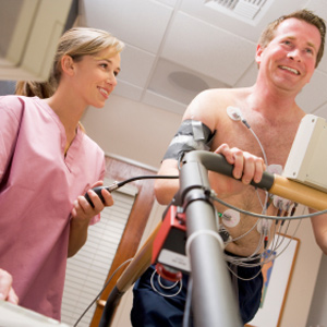
Exercise stress echocardiogram is a test performed to evaluate your heart’s function and pattern of heart rate during activity. It is indicated to check your heart’s tolerance to activity, assess the effectiveness of cardiac treatment and determine your chances of developing coronary artery disease (CAD).
